


Interfacial Stability Mechanisms at the Micro- and Nanoscale: From Nanobubbles to Nanodroplets
-
摘要: 微纳尺度气液与液液界面广泛存在于自然界与工程系统中, 其物理力学行为在尺度效应作用下显著不同于宏观体系. 纳米气泡与纳米液滴具有典型的软物质界面结构, 能够在极高拉普拉斯压强下长期稳定存在, 形成了著名的“稳定性悖论”: 对纳米气泡而言, 其长期稳定存在显著偏离爱泼斯坦–普莱塞特扩散模型的经典预测; 而对纳米液滴而言, 则表现出超出奥斯特瓦尔德熟化理论适用范围的非平衡稳定行为. 以连续介质力学与统计热力学为理论框架, 系统综述了表面纳米气泡、体相纳米气泡及纳米液滴的稳定性机理与演化规律. 重点分析了表面纳米气泡中接触线钉扎与界面气体过饱和对扩散过程的调制作用, 阐述了体相纳米气泡中界面电荷积累、离子特异性吸附及扩散屏蔽构建稳定性物理机制, 并总结了其对溶液环境及力学扰动的非线性响应特征. 进一步评述了多组分溶液体系中纳米液滴成核动力学及无表面活性剂微乳液中熵致的亚稳态稳定机制. 最后, 通过比较曲率效应、界面张力修正及界面分子排布, 构建了纳米气泡与纳米液滴的物理力学图像, 并展望了相关跨尺度建模、原位表征技术及能源环境领域的应用前景.Abstract: Micro- and nanoscale gas–liquid and liquid–liquid interfaces are ubiquitous in natural and engineering systems, where their physicomechanical behaviors differ significantly from those of macroscopic systems due to pronounced scale effects. Nanobubbles and nanodroplets represent typical soft-matter interfacial systems that can maintain long-term stability under extremely high Laplace pressures, leading to long-standing stability paradoxes. For nanobubbles, their persistent existence deviates significantly from the classical predictions of the Epstein–Plesset diffusion model, whereas nanodroplets exhibit nonequilibrium stabilization beyond the applicability of Ostwald ripening theory. Within the frameworks of continuum mechanics and thermodynamics, this review systematically summarizes the stability mechanisms and evolutionary behaviors of surface nanobubbles, bulk nanobubbles, and nanodroplets. The roles of contact-line pinning and interfacial gas supersaturation in regulating gas diffusion from surface nanobubbles are discussed, together with the stabilization mechanisms of bulk nanobubbles arising from interfacial charge accumulation, ion-specific adsorption, and diffusion shielding. The responses of nanobubble systems to variations in solution conditions and mechanical perturbations are further reviewed. In addition, the nucleation dynamics of nanodroplets in multicomponent solutions and the entropy-driven metastable stabilization mechanisms of surfactant-free microemulsions are discussed. Finally, by comparing curvature effects, interfacial tension corrections, and molecular arrangements at interfaces, a unified physicomechanical framework connecting nanobubbles and nanodroplets is established. Perspectives on multiscale modeling, in situ characterization techniques, and applications in energy and environmental fields are also provided.
-
Key words:
- nanobubbles /
- nanodroplets /
- gas-liquid interface /
- stability /
- micro/nanofluidics
-
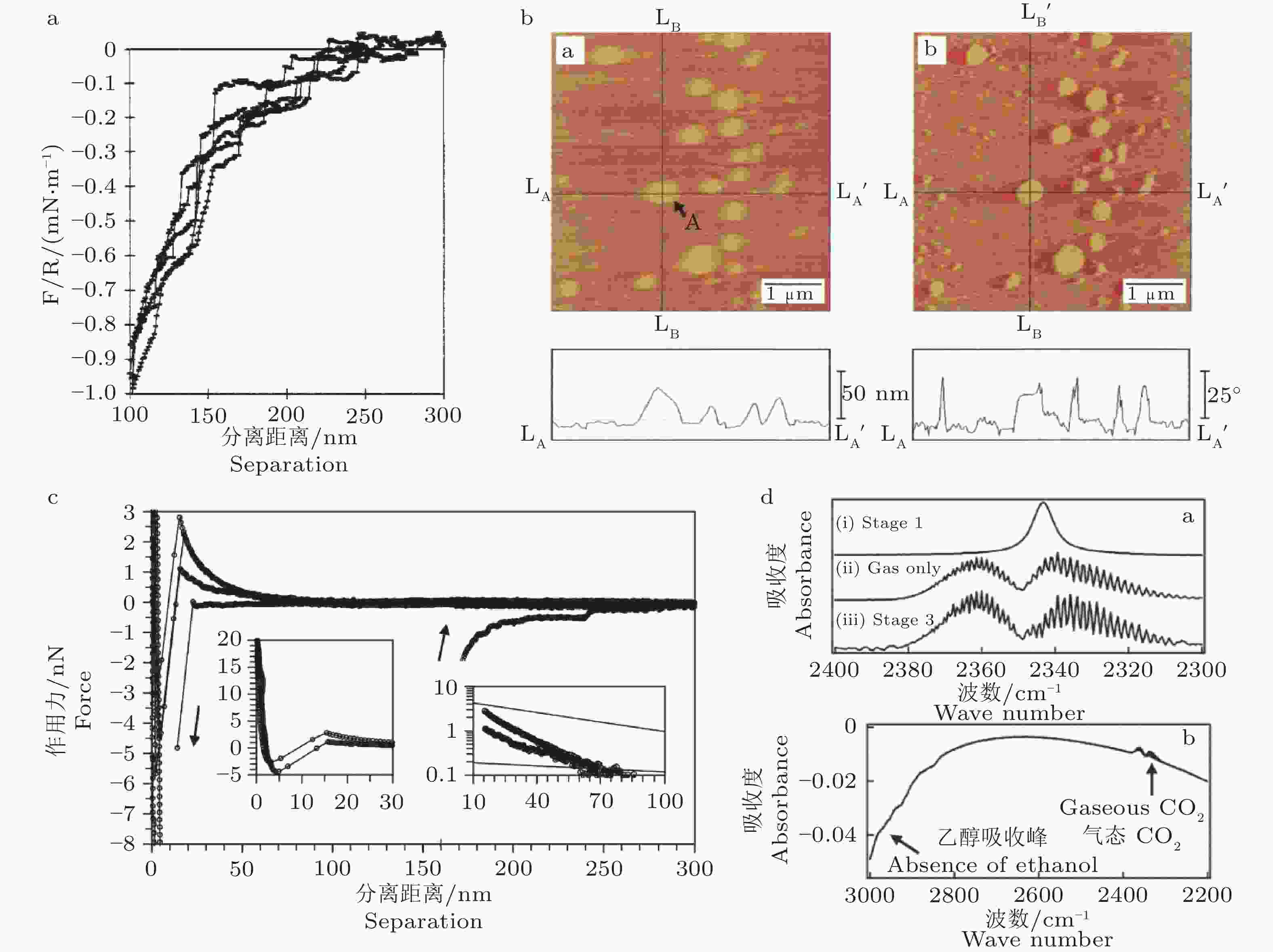
图 2 表面纳米气泡的多尺度表征. (a) 表面力曲线出现离散台阶特征, 被解释为固–液界面纳米气泡的生成与破裂过程(Parker et al. 1994). (b) 溶剂置换法在水–云母界面形成的表面纳米气泡形貌, A 指示典型单个气泡(Ishida et al. 2000). (c) 原子力显微镜测得力–距离曲线: 接近与回撤过程存在明显滞回, 回撤阶段跃迁表征探针与纳米气泡的黏附/脱附行为(Chan et al. 2012). (d) 衰减全反射傅里叶变换红外光谱显示气相 CO2 的转动精细结构, 证明乙醇–水置换后疏水硅–水界面存在纳米尺度气相, 并排除溶解态 CO2 与乙醇残留干扰 (Zhang X H et al. 2008).
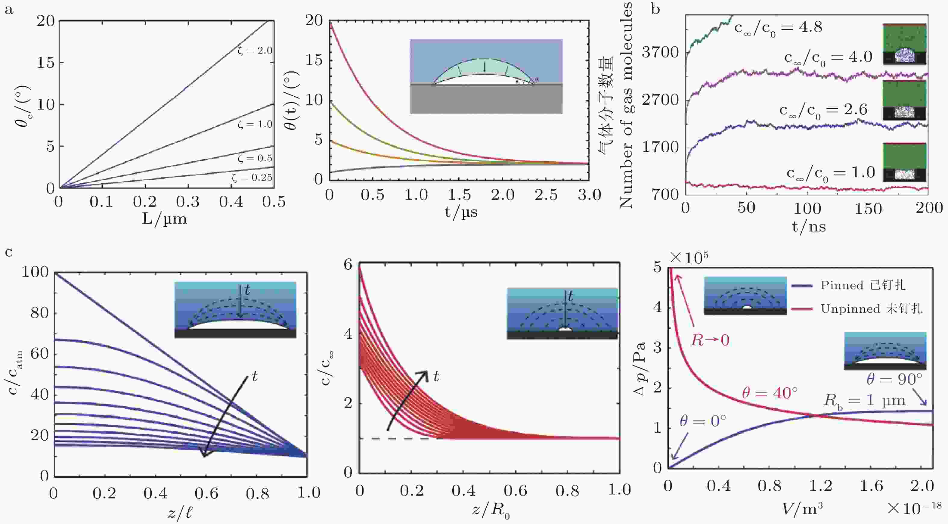
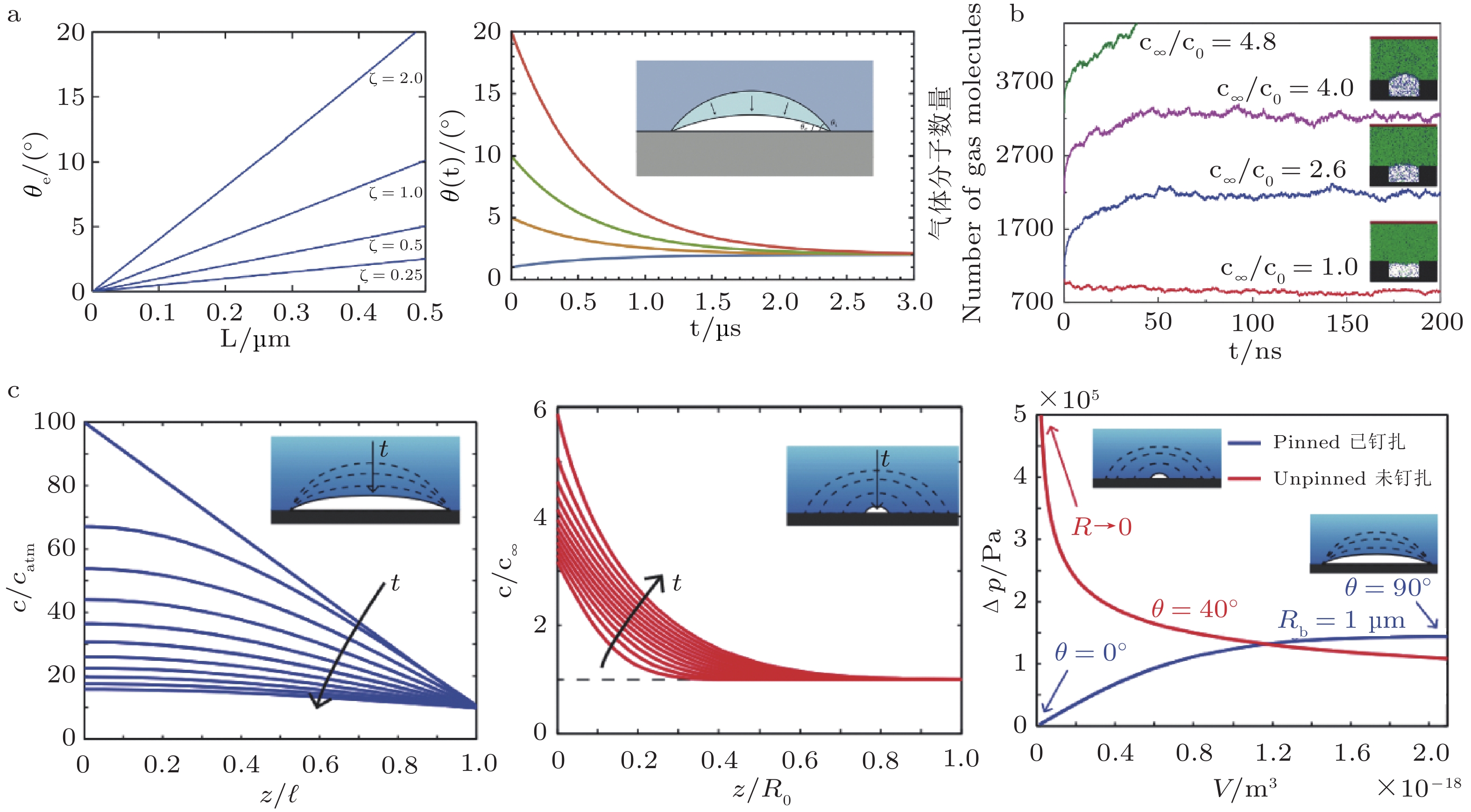
图 3 钉扎接触线条件下表面纳米气泡的扩散稳定性机理. (a) 在三相接触线被表面化学或几何非均匀性钉扎的情况下, 表面纳米气泡的体积演化由气体的扩散通量决定: 气泡内部较高的拉普拉斯压强驱动气体向外扩散, 而溶液中过饱和气体则向气泡内扩散, 当两者达到平衡时, 气泡可处于动力学稳定状态(Lohse et al. 2015b). (b) 在接触线钉扎条件下, 表面纳米气泡的接触角不再由 Young 方程决定, 而是由溶液的气体过饱和度确定, 过饱和度越高, 对应的稳定接触角越大(Liu et al. 2014). (c) 表面纳米气泡的稳定性相图, 显示只有在特定气体过饱和度与气泡尺寸范围内, 气泡才能实现扩散动力学稳定; 过小气泡因拉普拉斯压强过大而迅速溶解, 而过大气泡则无法维持扩散平衡(Lohse et al. 2015a).
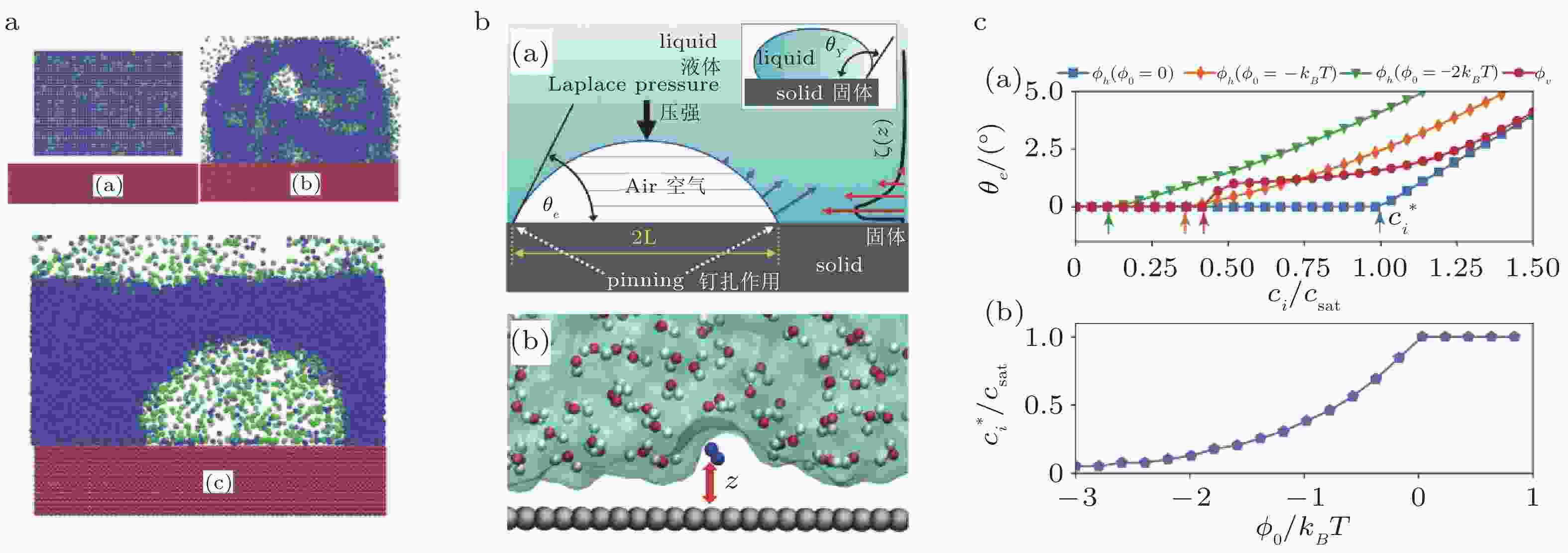
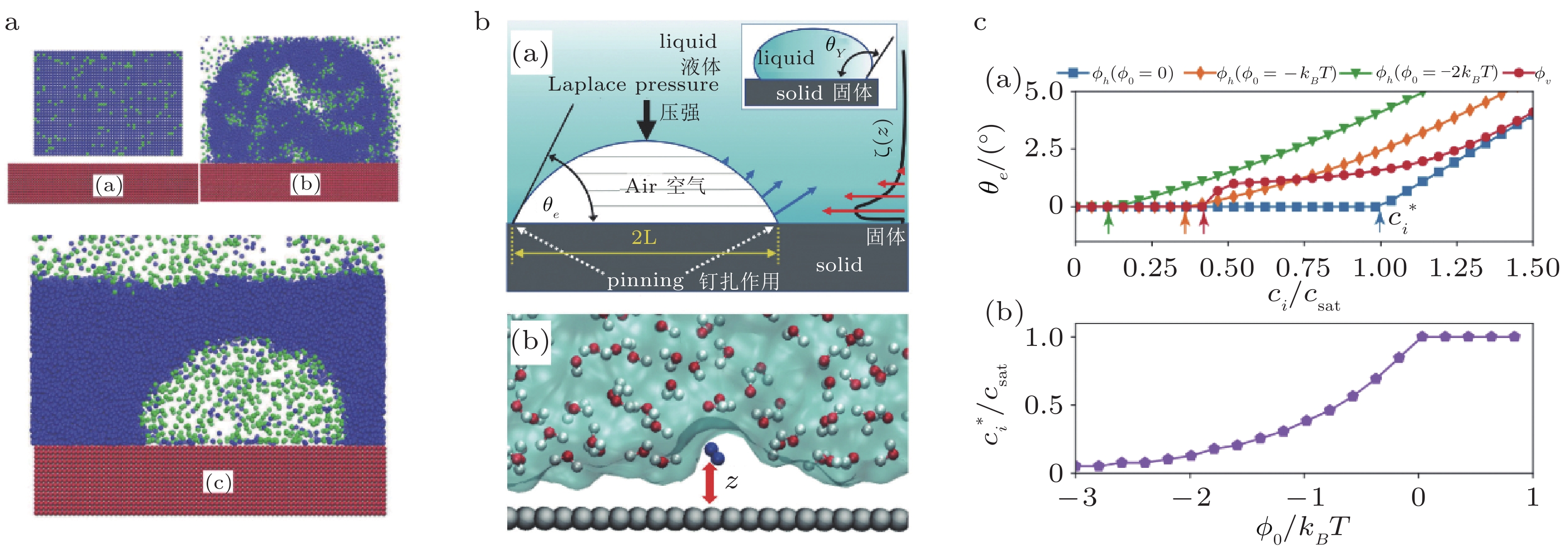
图 4 表面纳米气泡的形成、稳定机制及欠饱和环境响应. (a) 分子动力学模拟显示: 含气液膜覆盖基底后, 界面约 0.1 ns 成核、约 10 ns 形成稳定表面纳米气泡(Brenner et al. 2008). (b) 纳米气泡因三相接触线钉扎形成平衡接触角θe; 近壁区 ( ~ 1–2 nm) 局部过饱和补偿底部强外扩散通量, 使其在高拉普拉斯压力下仍可稳定(Tortora et al. 2020). (c) 平衡接触角随溶解气体浓度变化, 并受短程疏水吸引与范德华作用调控; 疏水性增强提高欠饱和耐受性, 而亲水或无疏水吸引时难以稳定(Tan et al. 2019).
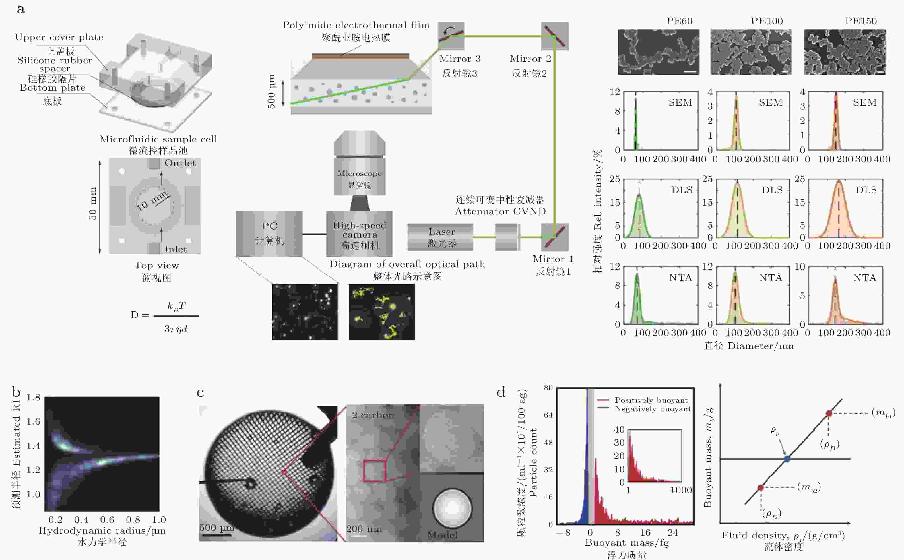
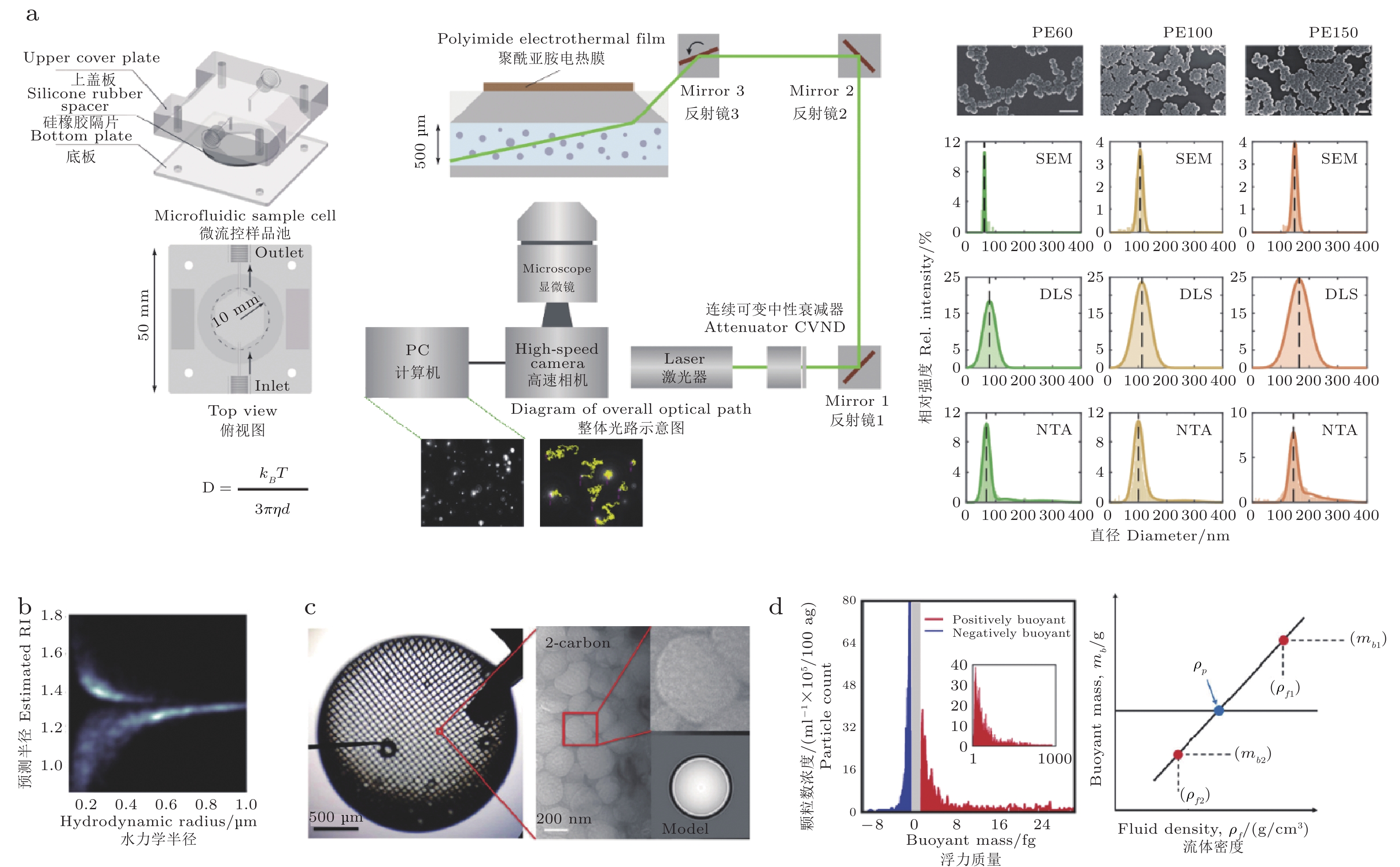
图 5 体相纳米气泡的多尺度表征. (a) 纳米颗粒跟踪系统装置图与两种检测技术NTA 与 DLS 对同一样品的粒径/浓度表征对比(Ma X T, Li, Sun, 2022). (b) H-NTA 联合反演亚波长目标的尺寸与折射率: 随粒径增大, 气泡/颗粒的估算折射率均趋近水 (n≈1.33), 但其光学响应差异可作为识别依据(Midtvedt et al. 2020). (c) 冷冻电镜直接成像体相纳米气泡, 为光学与力学测量提供独立的结构证据(Li M et al. 2016). (d) 基于平均浮力质量的密度测定: 在不同流体密度下测量浮力质量并外推至零浮力点以获得平均密度, 用于区分低密度气泡与固体/液体纳米颗粒(Alheshibri et al. 2018).
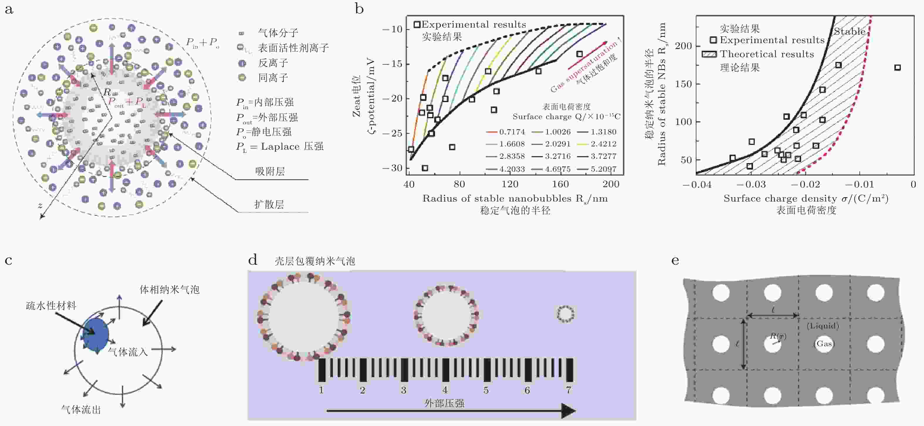
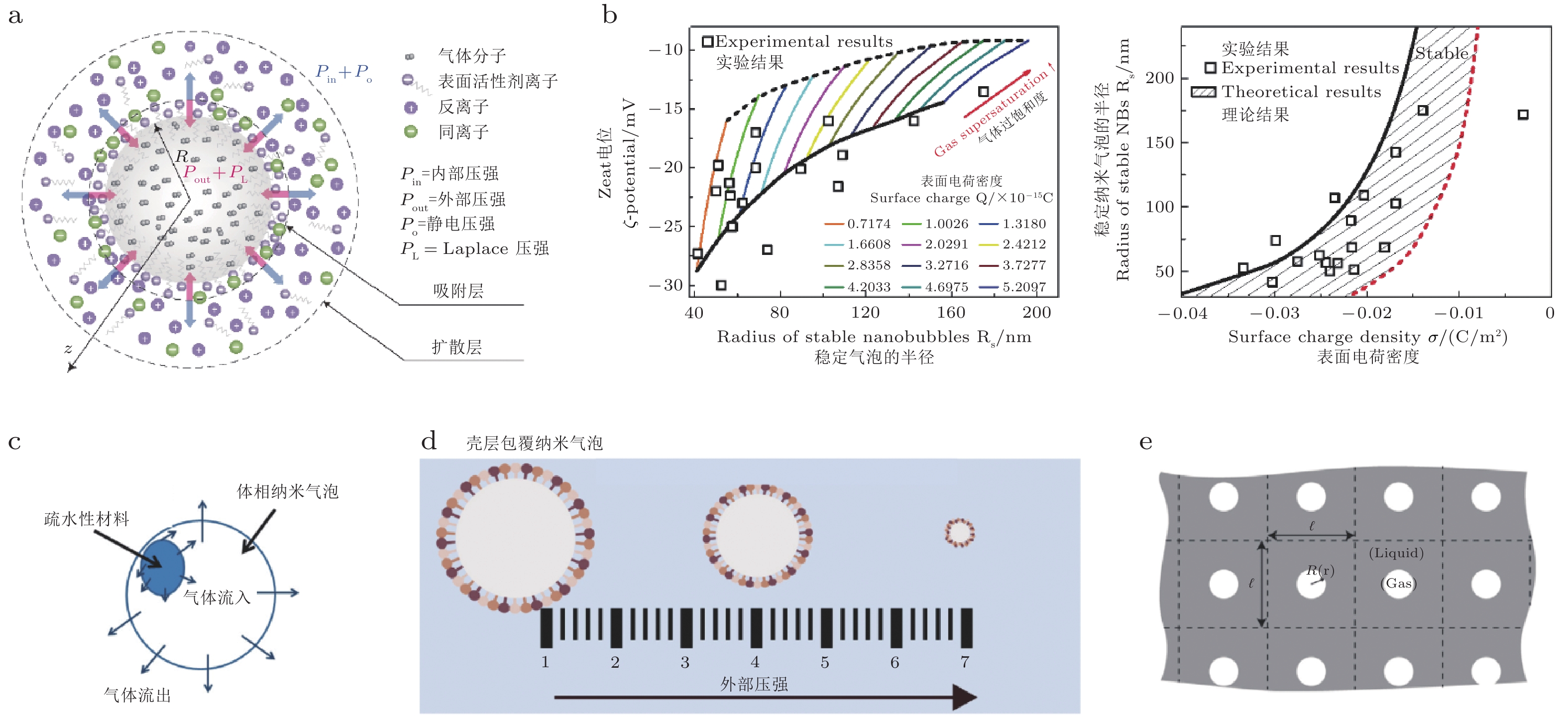
图 6 体相纳米气泡稳定性的典型机制. (a) 电荷稳定模型: 气泡界面离子富集形成扩散双电层, 界面电荷/电位改变气泡的有效稳定条件(Ma X et al. 2022). (b) 自由能模型与实验对照: 给出气泡半径与ζ电位/表面电荷密度的关系及稳定边界, 阴影区为由实验估计电荷密度范围限定的可稳定存在参数域(Zhang H G et al. 2020). (c) 疏水材料局部覆盖的体相气泡/微气泡: 覆盖改变界面边界条件与气体通量, 可能形成动态平衡式稳定(Yasui et al. 2016). (d)“装甲”纳米气泡: 界面被不溶性层/颗粒包覆, 在外加压力下表现出增强的结构稳定性(Alheshibri et al. 2019b). (e) 气泡簇集晶格模型: 相邻气泡间距ℓ决定扩散场相互作用; 小间距导致扩散屏蔽, 降低溶解驱动力并提高簇集稳定性(Weijs et al. 2012).
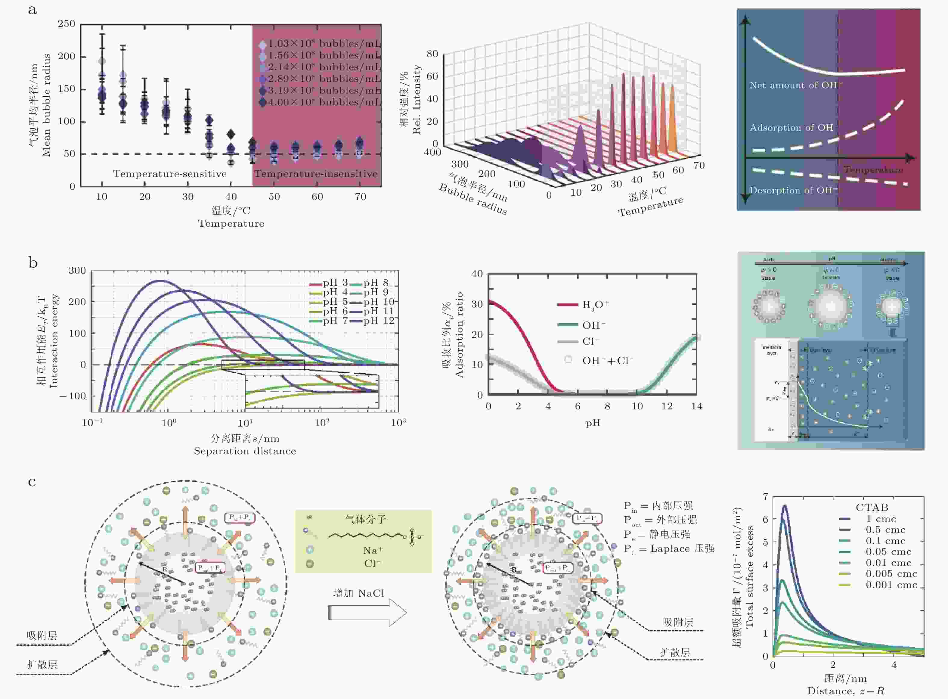
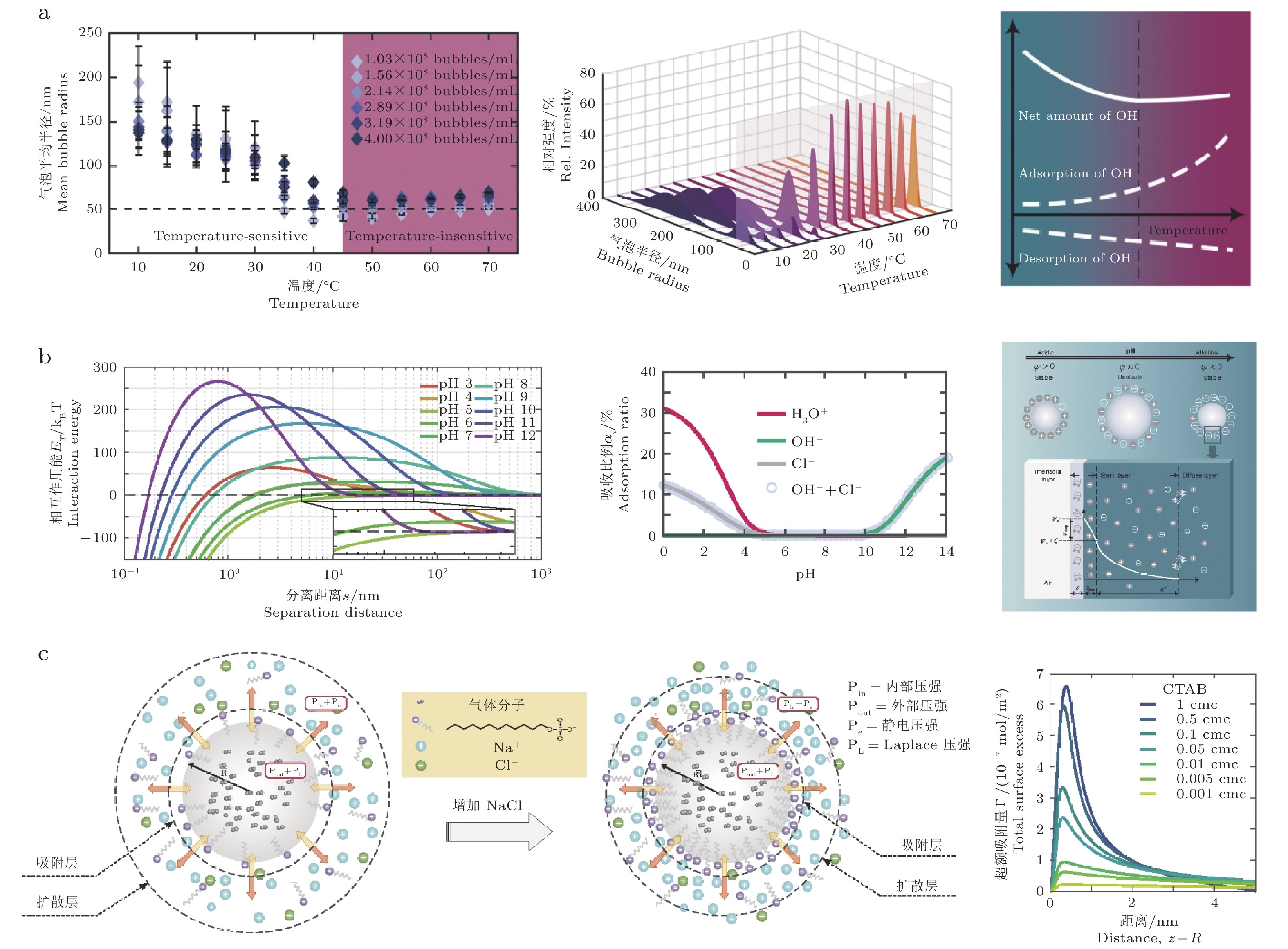
图 7 体相纳米气泡稳定性对环境因素的响应. (a) 温度效应: 随温度升高, 气泡粒径分布与平均半径$ {R}_{m} $发生系统变化, 并出现温度敏感/非敏感区. 该行为与气−液界面负电荷的积累有关, 其本质由 OH− 吸附与解吸两种竞争过程共同决定(Li M B et al. 2021). (b) pH效应: 不同 pH下离子 (H3O+、Cl−、OH−) 在气泡界面的吸附比例改变, 进而调控气泡表面电荷与扩散双电层结构; 由此 DLVO 相互作用势随 pH 改变, 影响纳米气泡的聚并/稳定区间(Ma X et al. 2022). (c) 离子强度/盐效应: NaCl 提高溶液离子强度并压缩电双层 (降低德拜长度), 改变电双层内离子分布与静电排斥, 从而调制气泡间相互作用、成核与长期稳定性 (通常表现为ζ电位与浓度/尺寸统计的改变)(Ma X T et al. 2024).
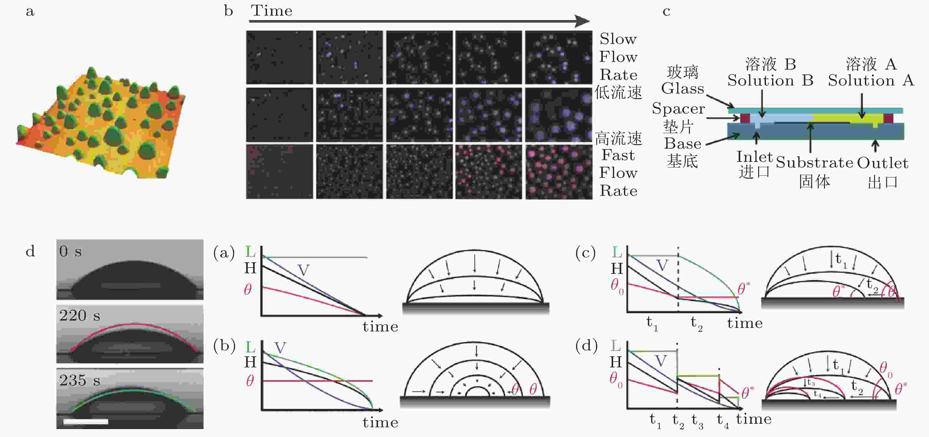
图 8 表面纳米液滴表征与动力学: 从形貌、生长到溶解模式. (a) AFM 表征的由溶剂置换法在疏水化硅表面生成的表面纳米液滴形貌 (30 × 30 μm2, 高度色标 0 ~ 800 nm), 显示其典型球冠几何与空间分布(Lohse et al. 2015a). (b) TIRF 原位快照显示溶剂置换过程中液滴随时间的成核与生长, 并对比不同流量 (25、35、50 μL·min−1) 下的生长速率及尺寸分布差异(Dyett et al. 2018). (c) 溶剂置换流道与窄通道几何 (h = 0.33 mm) 示意图, 通过控制流量调节界面过饱和脉冲, 从而控制液滴尺度与均匀性(Zhang X H, Lu, et al. 2015). (d) 表面纳米液滴的stick–jump溶解模式示意: 液滴先经历恒底径收缩使接触角降低至阈值θ*, 随后瞬时跳跃到新的几何构型, 直至最终消失(Dietrich et al. 2015; Zhang X H, Wang, et al. 2015).
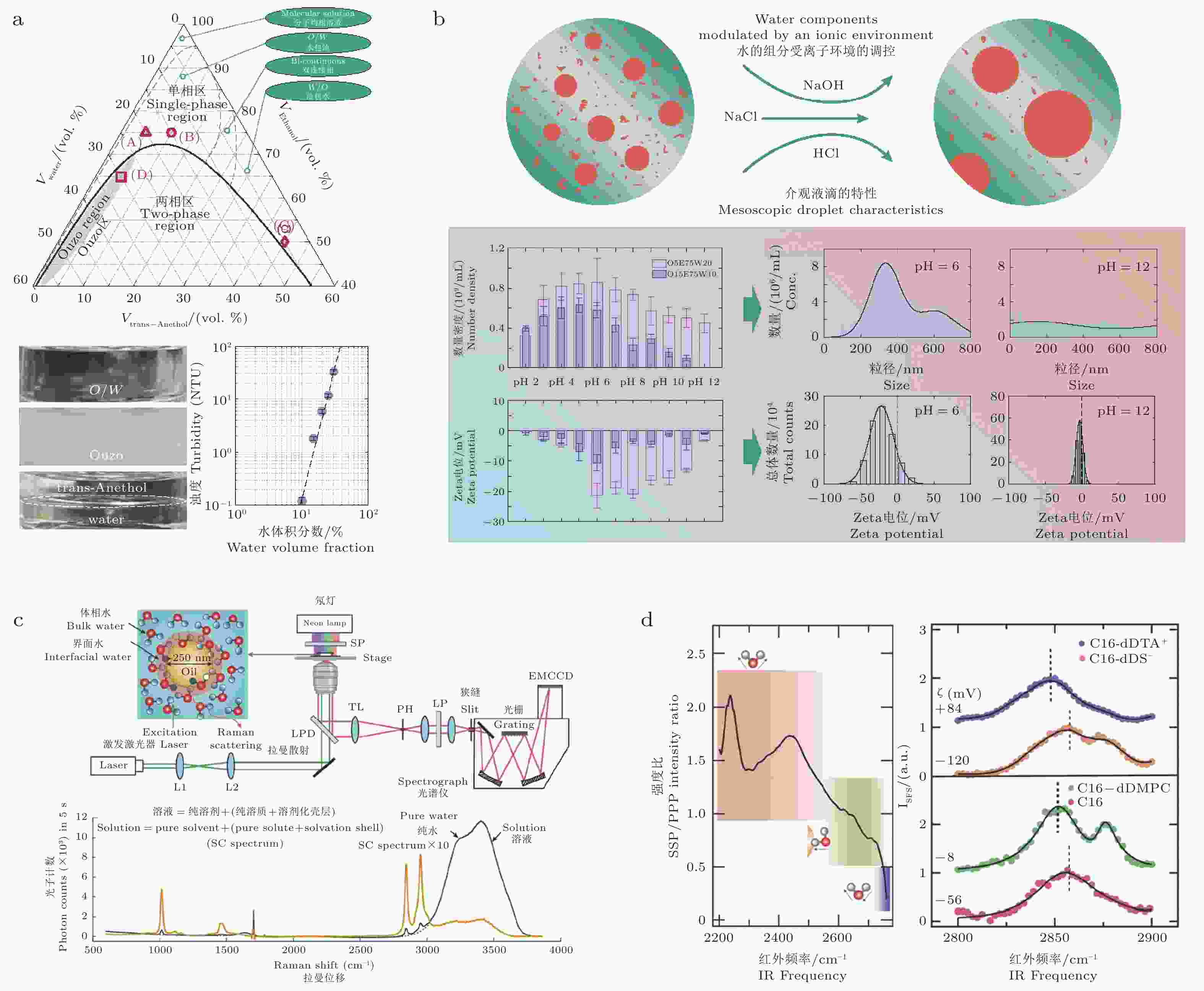
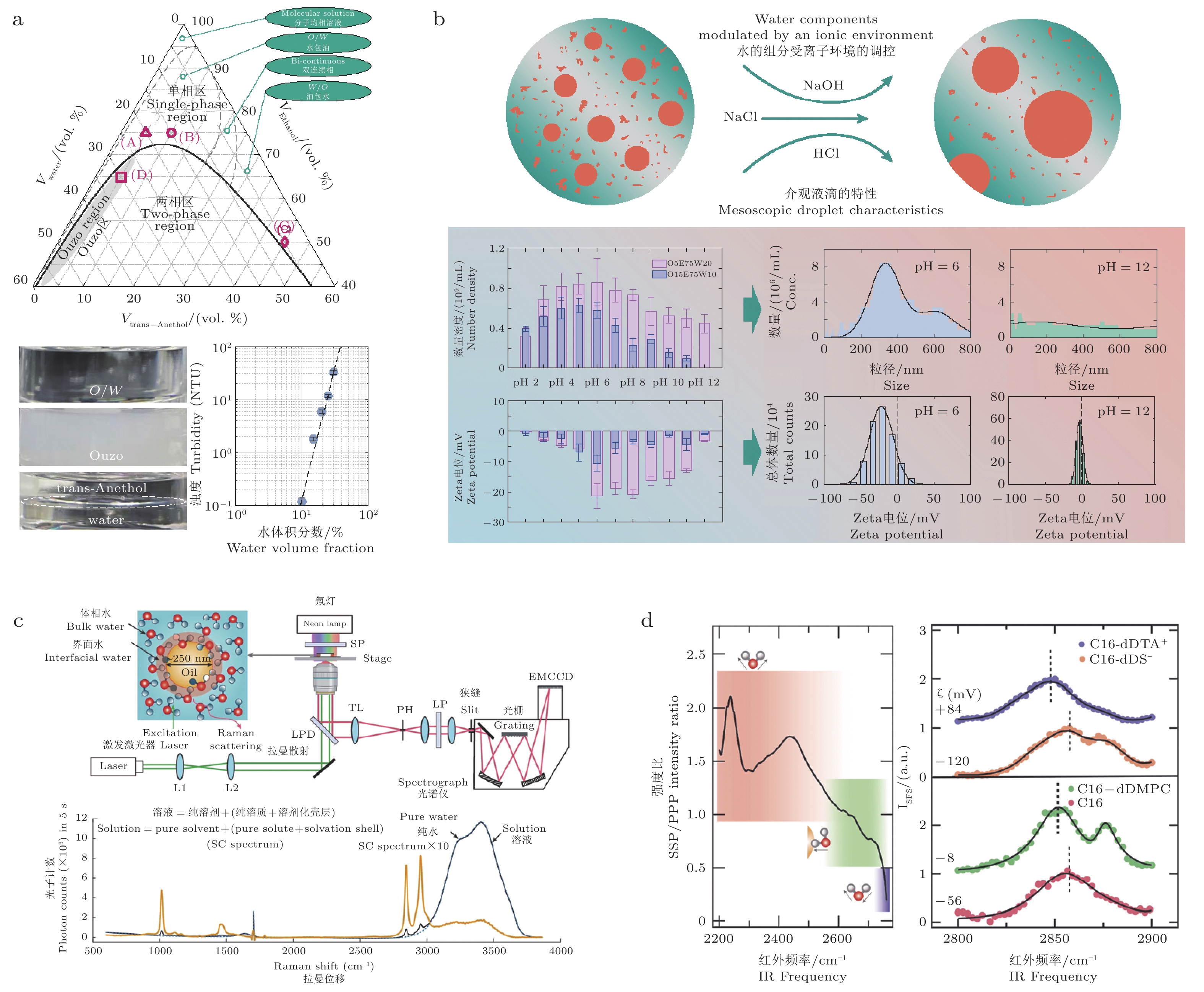
图 9 体相液滴/纳米域的相行为、离子环境调控与界面电学证据. (a) trans-anethol/乙醇/水三元体系 (25 ℃) 相图及沿组成路径进入 pre-Ouzo→Ouzo区域时的外观与浊度变化, 用于界定体相液滴/纳米域的生成窗口(Li M B et al. 2025). (b) 极端 pH 与离子强度改变 O/W 结构中纳米域的成核与长大: 液滴变大、数密度降低; 长大以 Ostwald 熟化为主(Gao Y W et al. 2025). (c) SFS 偏振与 C–H 光谱: O–D 偏振比与 C–H 峰位随界面电性 (ζ电位/表面覆盖) 变化, 指示界面电场/电荷转移对油滴稳定性的作用(Pullanchery et al. 2021). (d) 共焦微−拉曼装置示意, 用于原位测量油滴/水界面附近的水结构与电场相关光谱信息(Shi L X et al. 2025)
表 1 表面纳米气泡常用表征技术的空间/时间分辨率、优势与局限性
表征技术 典型空间/时间分辨率 主要优势 局限性 轻敲模式 AFM 垂向分辨率可达亚纳米级; 横向分辨率通常为数纳米至数十纳米; 单幅扫描通常为秒至分钟量级 可直接获得表面纳米气泡的高度、底径、形貌和表观接触角, 是确认其扁平球冠状几何特征的核心手段 探针可能扰动或压缩软气液界面; 横向尺寸受针尖卷积影响; 扫描速度较慢, 难以捕捉快速动力学过程 PeakForce QNM/
力谱 AFM空间分辨率与 AFM 相近; 力学响应分辨率可达 pN–nN 量级; 时间分辨率通常为
秒至分钟可同时获得形貌、黏附、耗散、变形和表观刚度信息, 有助于区分软气泡界面与硬质污染物 测得的是探针−界面耦合后的表观力学响应, 依赖加载力、扫描参数和模型假设; 仍可能扰动气泡 FM-AFM/非接触 AFM 垂向分辨率可达亚纳米甚至原子级; 时间分辨率通常较低 探针扰动较小, 可用于重构气液界面精细形貌, 判断界面是否具有高光滑度 技术门槛高, 成像区域有限; 不适合大范围统计; 对实验环境和反馈控制要求高 TIRF/TIRM 横向分辨率约200 ~ 300 nm; 倏逝场穿透深度约100 ~
200 nm; 时间分辨率可达
毫秒至秒量级可原位、非接触地观察近壁区域纳米气泡的成核、并合、溶解和周围颗粒运动, 对动态过程更敏感 主要探测近壁区域, 难以获得完整三维形貌; 通常依赖荧光或散射对比; 小于衍射极限的对象需要间接识别 SPR 表面等离子
激元共振对界面附近折射率变化高度敏感; 横向分辨率通常为微米量级; 时间分辨率可达
毫秒至秒量级可实时监测溶剂交换过程中气泡成核、生长和表面覆盖率变化, 适合研究界面平均动力学 不能直接给出单个气泡的三维形貌; 信号是折射率变化的综合响应, 需结合模型或其他成像方法解释 ATR-FTIR/
红外光谱空间分辨率通常较低; 倏逝波探测深度为亚微米至微米量级; 时间分辨率通常为秒至分钟 可提供气泡内部物质的化学证据, 例如识别气态CO2的特征红外吸收, 从而辅助排除聚合物污染或溶剂残留 主要提供化学平均信息, 不能直接给出单个气泡形貌; 适用体系受气体红外活性、基底和光谱灵敏度限制 光学显微/ 高速
显微成像横向分辨率受衍射极限限制, 通常约200 ~ 300 nm; 时间分辨率可达微秒至毫秒 适合观察较大气泡、气泡群和电极表面气泡成核/脱附动力
学; 可实现原位实时观测难以直接分辨典型表面纳米气泡的纳米高度和小尺寸结构; 对百纳米以下对象主要提供间接证据  下载: 导出CSV
表 2 表面纳米气泡主要稳定模型的适用条件、预测能力及局限性对比
稳定性模型 核心物理机制 适用条件 能解释的实验现象 局限性 接触线钉扎-
气体过饱和
模型接触线钉扎提供几何负反馈, 气体过饱和提供物质补偿; 二者共同使气泡达到稳定半径和接触角 基底存在粗糙度、化学异质性或缺陷位点; 液体中存在一定气体
过饱和可解释表面纳米气泡固定 footprint、异常小接触角、长寿命以及接触角随溶解过程变化等现象, 是目前最具定量预测能力的模型之一 需要明确钉扎边界和气体过饱和度; 单独的钉扎不能在欠饱和体系中长期稳定气泡 动态平衡 /
近壁富气层
模型疏水固体表面附近形成局部富气层或持续气体补给通道, 使纳米气泡的气体外扩散与近壁气体回流/补充达到动态平衡 疏水基底附近存在局部富气层, 或体系中存在持续气体补给 可解释气泡寿命远长于经典扩散理论预测值, 以及纳米气泡更易存在疏水表面
的现象气体回流或近壁富气层难以直接测量, 定量预测能力有限 污染膜 /
软钉扎模型痕量有机物、表面活性剂或高分子污染物吸附于气液界面, 形成低渗透性或黏弹性界面膜, 降低气体扩散通量 界面或基底存在表面活性剂、有机污染物、高分子吸附层或软物
质覆盖可解释部分实验中气泡界面“硬化”、气体渗透减慢、接触线缓慢移动等现象 容易与污染物、液滴或其他软物质结构混淆; 对洁净体系的普适
性有限界面电荷 /
双电层调控
模型气液界面吸附离子或形成带电界面, 电荷调控气泡间相互作用、接触线行为和界面自由能 pH、离子强度或表面活性剂显著影响气泡稳定性的体系 可解释气泡间排斥、聚并受抑制, 以及纳米气泡对盐浓度和 pH 的敏感性 通常只能作为辅助稳定机制; 难以单独抵消纳米气泡巨大的拉普
拉斯压力
下载: 导出CSV
表 3 体相纳米气泡常用表征技术的空间/时间分辨率、优势与局限性
表征技术 典型空间/时间分辨率 主要优势 主要局限性 DLS 动态光散射 可测水动力学粒径通常为纳米至微米; 时间分辨率通常
为秒至分钟操作简便、统计量大, 可快速获得体相分散体的平均粒径
和尺寸分布对大颗粒和少量污染物极其敏感; 多分散体系解析能力有限; 不能区分气泡、液滴、胶束或固体颗粒 NTA 纳米颗粒跟踪分析 通常适用于约20 nm–1 μm的散射体; 时间分辨率由视频帧率决定, 通常为毫秒至秒 可追踪单颗粒布朗运动, 获得粒径分布和数浓度; 相比DLS更适合多分散体系 依赖光散射对比, 低折射率小气泡检测困难; 仍不能单独证明
被测对象为气泡ζ电位/激光多普勒电泳 不提供直接形貌分辨率; 测量时间通常为分钟量级 可表征体相纳米气泡样分散体的界面带电性, 分析 pH、离子强度和表面活性剂对稳定性的影响 只能说明被测对象具有电泳迁移率或界面电性, 不能单独证明其为气泡; 液滴、胶束和颗粒也可能
具有类似ζ电位RMM 共振质量测量 单颗粒浮力质量测量; 典型适用于几十纳米到微米; 时间分辨率依赖于液体流速 可通过浮力质量符号区分低密度气泡样对象与高密度颗粒/液滴, 是体相纳米气泡相态鉴别
中的重要方法设备要求高, 通量和样品兼容性有限; 对极小、低浓度或易变形对象存在检测限制; 结果需结合标准样和控制实验解释 cryo-TEM/
cryo-EM空间分辨率可达纳米甚至亚纳米; 时间上为快速冻结后的
静态快照可直接观察纳米尺度低电子密度空腔、液滴或胶体结构, 为形貌证据提供重要补充 制样过程可能引入冻结、脱气、浓缩或空穴伪影; 视野小、统计量有限; 不是原位动态测量 DHM/H-NTA/iNTA 横向分辨率通常为亚微米至微米量级; 时间分辨率可达
视频帧率可实现非接触三维追踪, 并通过散射、相位或折射率信息辅助区分低折射率对象与固体颗粒 对更小、低折射率对比的纳米气泡检测困难; 相态判别依赖光学模型、反演算法或训练数据; 仍需交叉验证 SAXS/SANS小角散射 通常解析约1 ~ 100 nm尺度结构; 时间分辨率通常
为秒至分钟可获得体相平均结构、尺寸分布和界面密度差异信息, 适合研究纳米尺度分散结构的统计特征 模型依赖性强; 不同低密度或高密度纳米结构可能产生相似散射响应; 难以单独确认气泡相态 压力扰动/脱气/冷冻−融化对照 不提供直接空间分辨率; 时间尺度通常为分钟至小时 可通过环境响应验证气泡样对象是否随压力、溶解气浓度或脱气处理发生可逆/不可逆变化, 排除非气态颗粒的重要辅助证据 属于间接验证, 不能单独提供尺寸、形貌或内部相态信息; 需要与DLS/NTA/RMM/cryo-EM
等方法联用
下载: 导出CSV
表 4 体相纳米气泡主要稳定模型的适用条件、预测能力及局限性对比
稳定性模型 核心物理图像 适用条件 能解释的实验现象 局限性 电荷稳定模型 界面电荷产生的静电压强平衡表面张力引起的附加压强, 通过产生气泡间静电排斥, 抑制聚并, 并可能改变界面自由能 具有显著 ζ 电位, 且稳定性受 pH、离子强度
或表面活性剂影响可解释气泡带负电、气泡间静电排斥、聚并受抑制以及 ζ 电位、气泡尺寸分布与数量密度随溶液环境变化等现象 静电作用通常不足以完全平衡拉普拉斯压力 污染膜/
铠甲气泡模型痕量有机物、表面活性剂或高分子在气液界面吸附, 形成低渗透性或具有黏弹性的界面膜、颗粒壳层, 降低气体传质速率和气泡收缩 体系中存在表面活性剂、有机物、胶体颗粒
或固体杂质可解释气泡界面渗透性降低、寿命延长、对剪切和储存条件表现出较强稳定性等现象 容易引入污染伪影; 部分纳米粒子可能实际为液滴、颗粒或复合
胶体结构过饱和/
动态平衡模型气泡内部气体不断向外扩散, 同时环境中存在持续气体补给, 使气体流出与流入达到动态平衡. 体系中存在气体过饱和、持续气体生成、减压、电解、超声或温度扰动
等非平衡过程可解释体相纳米气泡的生成以及在一定时间内维持相对稳定的尺寸和浓度 需要明确持续气体来源; 对封闭、静止且无外部补气的体系解释
力较弱扩散屏蔽/
群体效应模型高浓度纳米气泡之间的扩散场相互重叠, 使单个气泡的气体逸散受到邻近气泡抑制, 从而延长整体寿命 纳米气泡浓度较高, 气泡间距较小, 扩散场
发生重叠可解释高数密度气泡群比孤立气泡更稳定, 以及气泡浓度对寿命的影响 对低浓度或孤立体相纳米气泡解释有限; 需要准确测量气泡空间
分布和数密度纳米尺度界面修正模型 纳米尺度下界面曲率可能改变有效表面张力, 从而降低拉普拉斯压力和溶解驱动力 气泡尺寸接近分子尺度, 界面曲率、表面张力或
热涨落效应不可忽略从界面热力学角度修正Young–Laplace方程 Epstein–Plesset扩散预测 实验验证困难; 通常难以单独解释百纳米尺度气泡的超长寿命
下载: 导出CSV
表 5 表面纳米液滴四种可能的溶解/蒸发模式
模式 过程 特点 适用场景 CCR (恒定接触半径) 接触线被牢固钉扎, 液滴高度降低, 接触角减小. 钉扎作用极强 粗糙表面或强缺陷位点 CCA (恒定接触半径) 接触线自由回缩, 液滴形状保持自相似. 无钉扎作用 理想光滑表面 Stick-Slip (粘−滑) 接触线交替进行“钉扎−滑动−钉扎”. 周期性运动 具有周期性或随机缺陷的表面 Stick-Jump (粘−跳) 接触线长时间钉扎, 随后瞬间去钉扎并
发生极快速的形态重组.离散体积跃变 光滑表面上的纳米级钉扎点
下载: 导出CSV
-
[1] 张立娟, 方海平, 胡钧. 2018. 纳米气泡的科学之谜. 物理, 47(9): 574-583 (Zhang L J, Fang H P, Hu J. 2018. Scientific mysteries of nanobubbles. Physics, 47(9): 574-583). doi: 10.7693/wl20180906Zhang L J, Fang H P, Hu J. 2018. Scientific mysteries of nanobubbles. Physics, 47(9): 574-583 doi: 10.7693/wl20180906 [2] 张雪花, 胡钧. 2004. 固液界面纳米气泡的研究进展. 化学进展, 16(5): 673-681. (Zhang X H, Hu J. 2004. Nanobubbles at the solid/water interface. Progress in Chemistry, 16(5): 673-681).Zhang X H, Hu J. 2004. Nanobubbles at the solid/water interface. Progress in Chemistry, 16(5): 673-681 [3] Abkarian M, Subramaniam A B, Kim S H, et al. 2007. Dissolution Arrest and Stability of Particle-Covered Bubbles. Physical Review Letters, 99(18): 188301. doi: 10.1103/PhysRevLett.99.188301 [4] Agarwal K, Trivedi M, Nirmalkar N. 2022. Does salting-out effect nucleate nanobubbles in water: Spontaneous nucleation. Ultrasonics Sonochemistry, 82: 105860. doi: 10.1016/j.ultsonch.2021.105860 [5] Agarwal K, Trivedi M, Ohl C D, et al. 2023. On Nanobubble Dynamics under an Oscillating Pressure Field during Salting-out Effects and Its DLVO Potential. Langmuir, 39(15): 5250-5262. doi: 10.1021/acs.langmuir.2c03085 [6] Akulichev V. 1966. Hydration of ions and the vacitation resistance of water. Acoustical Physics, 12(2): 144-150. [7] Alheshibri M, Craig V S J. 2018. Differentiating between Nanoparticles and Nanobubbles by Evaluation of the Compressibility and Density of Nanoparticles. The Journal of Physical Chemistry C, 122(38): 21998-22007. doi: 10.1021/acs.jpcc.8b07174 [8] Alheshibri M, Craig V S J. 2019a. Generation of nanoparticles upon mixing ethanol and water; Nanobubbles or Not. Journal of Colloid and Interface Science, 542: 136-143. doi: 10.1016/j.jcis.2019.01.134 [9] Alheshibri M, Craig V S J. 2019b. Armoured nanobubbles; ultrasound contrast agents under pressure. Journal of Colloid and Interface Science, 537: 123-131. doi: 10.1016/j.jcis.2018.10.108 [10] Alheshibri M, Qian J, Jehannin M, et al. 2016. A History of Nanobubbles. Langmuir, 32(43): 11086-11100. doi: 10.1021/acs.langmuir.6b02489 [11] Aljabri N M, Shi N, Cavazos A. 2022. Nanoemulsion: An emerging technology for oilfield applications between limitations and potentials. Journal of Petroleum Science and Engineering, 208: 109306. doi: 10.1016/j.petrol.2021.109306 [12] An H J, Tan B H, Ohl C D. 2016. Distinguishing Nanobubbles from Nanodroplets with AFM: The Influence of Vertical and Lateral Imaging Forces. Langmuir, 32(48): 12710-12715. doi: 10.1021/acs.langmuir.6b02519 [13] An H, Liu G, Atkin R, et al. 2015. Surface Nanobubbles in Nonaqueous Media: Looking for Nanobubbles in DMSO, Formamide, Propylene Carbonate, Ethylammonium Nitrate, and Propylammonium Nitrate. ACS Nano, 9(7): 7596-7607. doi: 10.1021/acsnano.5b02915 [14] Angulo A, Van Der Linde P, Gardeniers H, et al. 2020. Influence of Bubbles on the Energy Conversion Efficiency of Electrochemical Reactors. Joule, 4(3): 555-579. doi: 10.1016/j.joule.2020.01.005 [15] Attard P. 2014. The stability of nanobubbles. The European Physical Journal Special Topics, 223(5): 893-914. doi: 10.1140/epjst/e2013-01817-0 [16] Azmi N A, Elgharbawy A A M, Motlagh S R, et al. 2019. Nanoemulsions: Factory for Food, Pharmaceutical and Cosmetics. Processes, 7(9): 617. doi: 10.3390/pr7090617 [17] Bao L, Spandan V, Yang Y T, et al. 2018. Flow-induced dissolution of femtoliter surface droplet arrays. Lab on a Chip, 18(7): 1066-1074. doi: 10.1039/C7LC01321C [18] Bashkatov A, Hossain S S, Mutschke G, et al. 2022. On the growth regimes of hydrogen bubbles at microelectrodes. Physical Chemistry Chemical Physics, 24(43): 26738-26752. doi: 10.1039/D2CP02092K [19] Bilotto P, Miano D, Celebi A T, et al. 2024. Removal of Nanoparticles by Surface Nanobubbles Generated via Solvent–Water Exchange: A Critical Perspective. Langmuir, 40(52): 27127-27136. doi: 10.1021/acs.langmuir.4c02862 [20] Block B J, Das S K, Oettel M, et al. 2010. Curvature dependence of surface free energy of liquid drops and bubbles: A simulation study. The Journal of Chemical Physics, 133(15): 154702. doi: 10.1063/1.3493464 [21] Bokman G T, Biasiori-Poulanges L, Lukić B, et al. 2023. High-speed x-ray phase-contrast imaging of single cavitation bubbles near a solid boundary. Physics of Fluids, 35(1): 013322. doi: 10.1063/5.0132104 [22] Brenner M P, Lohse D. 2008. Dynamic Equilibrium Mechanism for Surface Nanobubble Stabilization. Physical Review Letters, 101(21): 214505. doi: 10.1103/PhysRevLett.101.214505 [23] Bu X N, Alheshibri M. 2021. The effect of ultrasound on bulk and surface nanobubbles: A review of the current status. Ultrasonics Sonochemistry, 76: 105629. doi: 10.1016/j.ultsonch.2021.105629 [24] Bull D S, Nelson N, Konetski D, et al. 2018. Contact Line Pinning Is Not Required for Nanobubble Stability on Copolymer Brushes. The Journal of Physical Chemistry Letters, 9(15): 4239-4244. doi: 10.1021/acs.jpclett.8b01723 [25] Bunkin N F, Shkirin A V, Suyazov N V, et al. 2016. Formation and Dynamics of Ion-Stabilized Gas Nanobubble Phase in the Bulk of Aqueous NaCl Solutions. The Journal of Physical Chemistry B, 120(7): 1291-1303. doi: 10.1021/acs.jpcb.5b11103 [26] Caupin F, Herbert E. 2006. Cavitation in water: a review. Comptes Rendus. Physique, 7(9-10): 1000-1017. doi: 10.1016/j.crhy.2006.10.015 [27] Chan C U, Ohl C D. 2012. Total-Internal-Reflection-Fluorescence Microscopy for the Study of Nanobubble Dynamics. Physical Review Letters, 109(17): 174501. doi: 10.1103/PhysRevLett.109.174501 [28] Chang A S, Niu B, Liu J, et al. 2023. Enrichment of surface charge contributes to the stability of surface nanobubbles. Particuology, 81: 128-134. doi: 10.1016/j.partic.2023.01.001 [29] Chang C C, Sheng Y J, Tsao H K. 2016. Wetting hysteresis of nanodrops on nanorough surfaces. Physical Review E, 94(4): 042807. doi: 10.1103/PhysRevE.94.042807 [30] Chen C S, Gao Y W, Wang F, et al. 2025. A multiscale model for bubble nucleation thresholds on solid walls in the presence of nanometre-sized defects. Journal of Fluid Mechanics, 1015: A47. doi: 10.1017/jfm.2025.10374 [31] Chen C S, Gao Y W, Zhang X R. 2025. The Existence and Stability Mechanism of Bulk Nanobubbles: A Review. Nanomaterials, 15(4): 314. doi: 10.3390/nano15040314 [32] Chen Y Y, Hu Y, Wang B L, et al. 2024. Interfacial Thermal Fluctuations Stabilize Bulk Nanobubbles. Physical Review Letters, 133(10): 104001. doi: 10.1103/PhysRevLett.133.104001 [33] Collini H, Jackson M D. 2023. Zeta potential of crude oil in aqueous solution. Advances in Colloid and Interface Science, 320: 102962. doi: 10.1016/j.cis.2023.102962 [34] Creux P, Lachaise J, Graciaa A, et al. 2007. Specific cation effects at the hydroxide-charged air/water interface. The Journal of Physical Chemistry C, 111(9): 3753-3755. doi: 10.1021/jp070060s [35] Dammer S M, Lohse D. 2006. Gas Enrichment at Liquid-Wall Interfaces. Physical Review Letters, 96(20): 206101. doi: 10.1103/PhysRevLett.96.206101 [36] Dietrich E, Kooij E S, Zhang X H, et al. 2015. Stick-Jump Mode in Surface Droplet Dissolution. Langmuir, 31(16): 4696-4703. doi: 10.1021/acs.langmuir.5b00653 [37] Dollet B, Lohse D. 2016. Pinning Stabilizes Neighboring Surface Nanobubbles against Ostwald Ripening. Langmuir, 32(43): 11335-11339. doi: 10.1021/acs.langmuir.6b02136 [38] Duncan P B, Needham D. 2004. Test of the Epstein−Plesset Model for Gas Microparticle Dissolution in Aqueous Media: Effect of Surface Tension and Gas Undersaturation in Solution. Langmuir, 20(7): 2567-2578. doi: 10.1021/la034930i [39] Dyett B, Kiyama A, Rump M, et al. 2018. Growth dynamics of surface nanodroplets during solvent exchange at varying flow rates. Soft Matter, 14(25): 5197-5204. doi: 10.1039/C8SM00705E [40] Eftekhari-Bafrooei A, Borguet E. 2009. Effect of Surface Charge on the Vibrational Dynamics of Interfacial Water. Journal of the American Chemical Society, 131(34): 12034-12035. doi: 10.1021/ja903340e [41] Eklund F, Alheshibri M, Swenson J. 2021. Differentiating bulk nanobubbles from nanodroplets and nanoparticles. Current Opinion in Colloid & Interface Science, 53: 101427. doi: 10.1016/j.cocis.2021.101427 [42] Epstein P S, Plesset M S. 1950. On the stability of gas bubbles in liquid-gas solutions. The Journal of Chemical Physics, 18(11): 1505-1509. doi: 10.1063/1.1747520 [43] Factorovich M H, Molinero V, Scherlis D A. 2014. Vapor Pressure of Water Nanodroplets. Journal of the American Chemical Society, 136(12): 4508-4514. doi: 10.1021/ja405408n [44] Fang C K, Ko H C, Yang C W, et al. 2016. Nucleation processes of nanobubbles at a solid/water interface. Scientific Reports, 6(1): 24651. doi: 10.1038/srep24651 [45] Ferrara K, Pollard R, Borden M. 2007. Ultrasound microbubble contrast agents: fundamentals and application to gene and drug delivery. Annu. Rev. Biomed. Eng., 9(1): 415-447. doi: 10.1146/annurev.bioeng.8.061505.095852 [46] Ferraro G, Jadhav A J, Barigou M. 2020. A Henry’s law method for generating bulk nanobubbles. Nanoscale, 12(29): 15869-15879. doi: 10.1039/D0NR03332D [47] Filipe V, Hawe A, Jiskoot W. 2010. Critical evaluation of Nanoparticle Tracking Analysis (NTA) by NanoSight for the measurement of nanoparticles and protein aggregates. Pharmaceutical research, 27(5): 796-810. doi: 10.1007/s11095-010-0073-2 [48] Fischer V, Marcus J, Touraud D, et al. 2015. Toward surfactant-free and water-free microemulsions. Journal of Colloid and Interface Science, 453: 186-193. doi: 10.1016/j.jcis.2015.04.069 [49] Freedman M A, Huang Q S, Pitta K R. 2024. Phase transitions in organic and organic/inorganic aerosol particles. Annual Review of Physical Chemistry, 75. [50] Ganachaud F, Katz J L. 2005. Nanoparticles and Nanocapsules Created Using the Ouzo Effect: Spontaneous Emulsification as an Alternative to Ultrasonic and High-Shear Devices. ChemPhysChem, 6(2): 209-216. doi: 10.1002/cphc.200400527 [51] Gao Y W, Chen C S, Li M B, et al. 2025. Ionic environment-modulated nucleation and stability of multiscale nanodomains in surfactant-free microemulsions. Journal of Colloid and Interface Science, 696: 137833. doi: 10.1016/j.jcis.2025.137833 [52] Gao Z, Wu W X, Sun W T, et al. 2021. Understanding the Stabilization of a Bulk Nanobubble: A Molecular Dynamics Analysis. Langmuir, 37(38): 11281-11291. doi: 10.1021/acs.langmuir.1c01796 [53] German S R, Wu X, An H J, et al. 2014. Interfacial Nanobubbles Are Leaky: Permeability of the Gas/Water Interface. ACS Nano, 8(6): 6193-6201. doi: 10.1021/nn5016049 [54] Grapentin C, Barnert S, Schubert R. 2015. Monitoring the stability of perfluorocarbon nanoemulsions by Cryo-TEM image analysis and dynamic light scattering. PLoS One, 10(6): e0130674. doi: 10.1371/journal.pone.0130674 [55] Gray-Weale A, Beattie J K. 2009. An explanation for the charge on water’s surface. Physical Chemistry Chemical Physics, 11(46): 10994-11005. doi: 10.1039/b901806a [56] Gupta A, Eral H B, Hatton T A, et al. 2016. Nanoemulsions: formation, properties and applications. Soft Matter, 12(11): 2826-2841. doi: 10.1039/C5SM02958A [57] Hossain S S, Bashkatov A, Yang X, et al. 2022. Force balance of hydrogen bubbles growing and oscillating on a microelectrode. Physical Review E, 106(3): 035105. doi: 10.1103/physreve.106.035105 [58] Hou W G, Xu J. 2016. Surfactant-free microemulsions. Current Opinion in Colloid & Interface Science, 25: 67-74. doi: 10.1016/j.cocis.2016.06.013 [59] Ishida N, Inoue T, Miyahara M, et al. 2000. Nano Bubbles on a Hydrophobic Surface in Water Observed by Tapping-Mode Atomic Force Microscopy. Langmuir, 16(16): 6377-6380. doi: 10.1021/la000219r [60] Israelachvili J, Pashley R. 1982. The hydrophobic interaction is long range, decaying exponentially with distance. Nature, 300(5890): 341-342. doi: 10.1038/300341a0 [61] Jadhav A J, Barigou M. 2020. Bulk Nanobubbles or Not Nanobubbles: That is the Question. Langmuir, 36(7): 1699-1708. doi: 10.1021/acs.langmuir.9b03532 [62] Jadhav A J, Barigou M. 2021. Electrochemically Induced Bulk Nanobubbles. Industrial & Engineering Chemistry Research, 60(49): 17999-18006. doi: 10.1021/acs.iecr.1c04046 [63] Janek J, Zeier W G. 2016. A solid future for battery development. Nature Energy, 1(9): 16141. doi: 10.1038/nenergy.2016.141 [64] Johnson B D, Cooke R C. 1981. Generation of Stabilized Microbubbles in Seawater. Science, 213(4504): 209-211. doi: 10.1126/science.213.4504.209 [65] Ju L, Guo Z, Yan B, et al. 2024. Implementation of contact line motion based on the phase-field lattice Boltzmann method. Physical Review E, 109(4): 045307. [66] Kanduč M. 2017. Going beyond the standard line tension: Size-dependent contact angles of water nanodroplets. The Journal of Chemical Physics, 147(17): 174701. doi: 10.1063/1.4990741 [67] Kanduč M, Eixeres L, Liese S, et al. 2018. Generalized line tension of water nanodroplets. Physical Review E, 98(3): 032804. doi: 10.1103/PhysRevE.98.032804 [68] Kim J Y, Song M G, Kim J D. 2000. Zeta Potential of Nanobubbles Generated by Ultrasonication in Aqueous Alkyl Polyglycoside Solutions. Journal of Colloid and Interface Science, 223(2): 285-291. doi: 10.1006/jcis.1999.6663 [69] Koroleva M Y, Yurtov E V. 2021. Ostwald ripening in macro-and nanoemulsions. Russian Chemical Reviews, 90(3): 293. doi: 10.1070/RCR4962 [70] Leong K Y, Wang F. 2018. A molecular dynamics investigation of the surface tension of water nanodroplets and a new technique for local pressure determination through density correlation. The Journal of Chemical Physics, 148(14): 144503. doi: 10.1063/1.5004985 [71] Lepeltier E, Bourgaux C, Couvreur P. 2014. Nanoprecipitation and the “Ouzo effect”: Application to drug delivery devices. 2014 Editor’s Collection, 71: 86-97. [72] Li M B, Gao Y W, Ma X T, et al. 2024. How bulk nanobubbles respond to elevated external pressures. Physics of Fluids, 36(9). [73] Li M B, Lai R, Tian Y D, et al. 2025. Uncovering the Nanoscopic Phase Behavior of Ternary Solutions in the Presence of Electrolytes: From Pre-Ouzo to Ouzo Regions. Langmuir, 41(40): 27381-27392. doi: 10.1021/acs.langmuir.5c03507 [74] Li M B, Ma X T, Eisener J, et al. 2021. How bulk nanobubbles are stable over a wide range of temperatures. Journal of Colloid and Interface Science, 596: 184-198. doi: 10.1016/j.jcis.2021.03.064 [75] Li M B, Yi L, Sun C. 2022. Spontaneously formed multiscale nano-domains in monophasic region of ternary solution. Journal of Colloid and Interface Science, 628: 223-235. doi: 10.1016/j.jcis.2022.07.152 [76] Li M, Tonggu L, Zhan X, et al. 2016. Cryo-EM Visualization of Nanobubbles in Aqueous Solutions. Langmuir, 32(43): 11111-11115. doi: 10.1021/acs.langmuir.6b00261 [77] Li M, Wakata Y, Zeng H, et al. 2023. On the thermal response of multiscale nanodomains formed in trans-anethol/ethanol/water surfactant-free microemulsion. Journal of Colloid and Interface Science, 652: 1944-1953. doi: 10.1016/j.jcis.2023.08.166 [78] Li T, Cui Z, Sun J, et al. 2021. Generation of Bulk Nanobubbles by Self-Developed Venturi-Type Circulation Hydrodynamic Cavitation Device. Langmuir, 37(44): 12952-12960. doi: 10.1021/acs.langmuir.1c02010 [79] Lifshitz I M, Slyozov V V. 1961. The kinetics of precipitation from supersaturated solid solutions. Journal of Physics and Chemistry of Solids, 19(1): 35-50. doi: 10.1016/0022-3697(61)90054-3 [80] Liu Y W, Zhang X R. 2013. Nanobubble stability induced by contact line pinning. The Journal of Chemical Physics, 138(1): 014706. doi: 10.1063/1.4773249 [81] Liu Y W, Zhang X R. 2014. A unified mechanism for the stability of surface nanobubbles: Contact line pinning and supersaturation. The Journal of Chemical Physics, 141(13): 134702. doi: 10.1063/1.4896937 [82] Liu Y W, Zhang X R. 2017. Molecular dynamics simulation of nanobubble nucleation on rough surfaces. The Journal of Chemical Physics, 146(16): 164704. doi: 10.1063/1.4981788 [83] Lohse D, Zhang X H. 2015a. Surface nanobubbles and nanodroplets. Reviews of Modern Physics, 87(3): 981-1035. doi: 10.1103/RevModPhys.87.981 [84] Lohse D, Zhang X H. 2015b. Pinning and gas oversaturation imply stable single surface nanobubbles. Physical Review E, 91(3): 031003. doi: 10.1103/PhysRevE.91.031003 [85] Lou S T, Ouyang Z Q, Zhang Y, et al. 2000. Nanobubbles on solid surface imaged by atomic force microscopy. Journal of Vacuum Science & Technology B: Microelectronics and Nanometer Structures, 18(5): 2573. [86] Lu Z Y, Schaarsberg M H K, Zhu X J, et al. 2017. Universal nanodroplet branches from confining the Ouzo effect. Proceedings of the National Academy of Sciences, 114(39): 10332-10337. doi: 10.1073/pnas.1704727114 [87] Ma W, Du N, Hou W. 2025. Thermodynamic Explanation of Surfactant-Free Microemulsions. The Journal of Physical Chemistry B, 129(7): 2115-2127. doi: 10.1021/acs.jpcb.4c08376 [88] Ma X, Li M, Pfeiffer P, et al. 2022. Ion adsorption stabilizes bulk nanobubbles. Journal of Colloid and Interface Science, 606: 1380-1394. doi: 10.1016/j.jcis.2021.08.101 [89] Ma X T, Li M B, Sun C. 2022. Measurement and characterization of bulk nanobubbles by nanoparticle tracking analysis method. Journal of Hydrodynamics, 34(6): 1121-1133. doi: 10.1007/s42241-022-0077-5 [90] Ma X T, Li M B, Sun C. 2024. Effect of ionic environment in aqueous solution on nucleation and stabilization of bulk nanobubbles. Applied Surface Science, 656: 159726. doi: 10.1016/j.apsusc.2024.159726 [91] Ma X T, Li M B, Xu X, et al. 2023. On the role of surface charge and surface tension tuned by surfactant in stabilizing bulk nanobubbles. Applied Surface Science, 608: 155232. doi: 10.1016/j.apsusc.2022.155232 [92] Ma X T, Li M B, Xu X F, et al. 2022. Coupling Effects of Ionic Surfactants and Electrolytes on the Stability of Bulk Nanobubbles. Nanomaterials, 12(19): 3450. doi: 10.3390/nano12193450 [93] Maheshwari S, van der Hoef M, Rodrı́guez Rodrı́guez J, et al. 2018. Leakiness of Pinned Neighboring Surface Nanobubbles Induced by Strong Gas–Surface Interaction. ACS Nano, 12(3): 2603-2609. doi: 10.1021/acsnano.7b08614 [94] Maheshwari S, van der Hoef M, Zhang X H, et al. 2016. Stability of Surface Nanobubbles: A Molecular Dynamics Study. Langmuir, 32(43): 11116-11122. doi: 10.1021/acs.langmuir.6b00963 [95] Malm A V, Corbett J C W. 2019. Improved Dynamic Light Scattering using an adaptive andstatistically driven time resolved treatment of correlation data. Scientific Reports, 9(1): 13519. doi: 10.1038/s41598-019-50077-4 [96] Manning G S. 2020. On the thermodynamic stability of bubbles, immiscible droplets, and cavities. Physical Chemistry Chemical Physics, 22(31): 17523-17531. doi: 10.1039/D0CP02517H [97] Matyushov D V. 2014. Electrophoretic mobility without charge driven by polarisation of the nanoparticle–water interface. Molecular Physics, 112(15): 2029-2039. doi: 10.1080/00268976.2014.882521 [98] Meegoda J N, Aluthgun Hewage S, Batagoda J H. 2018. Stability of Nanobubbles. Environmental Engineering Science, 35(11): 1216-1227. doi: 10.1089/ees.2018.0203 [99] Meegoda J N, Hewage S A, Batagoda J H. 2019. Application of the Diffused Double Layer Theory to Nanobubbles. Langmuir, 35(37): 12100-12112. doi: 10.1021/acs.langmuir.9b01443 [100] Midtvedt D, Eklund F, Olsén E, et al. 2020. Size and Refractive Index Determination of Subwavelength Particles and Air Bubbles by Holographic Nanoparticle Tracking Analysis. Analytical Chemistry, 92(2): 1908-1915. doi: 10.1021/acs.analchem.9b04101 [101] Mørch K A. 2007. Reflections on cavitation nuclei in water. Physics of Fluids, 19(7): 072104. doi: 10.1063/1.2747210 [102] Nirmalkar N, Pacek A W, Barigou M. 2018. Interpreting the interfacial and colloidal stability of bulk nanobubbles. Soft Matter, 14(47): 9643-9656. doi: 10.1039/C8SM01949E [103] Nirmalkar N, Pacek A W, Barigou M. 2019. Bulk Nanobubbles from Acoustically Cavitated Aqueous Organic Solvent Mixtures. Langmuir, 35(6): 2188-2195. doi: 10.1021/acs.langmuir.8b03113 [104] Niwano M, Ma T, Iwata K, et al. 2023. Two-dimensional water-molecule-cluster layers at nanobubble interfaces. Journal of Colloid and Interface Science, 652: 1775-1783. doi: 10.1016/j.jcis.2023.08.173 [105] Park S, Liu L H, Demirkır Ç, et al. 2023. Solutal Marangoni effect determines bubble dynamics during electrocatalytic hydrogen evolution. Nature Chemistry, 15(11): 1532-1540. doi: 10.1038/s41557-023-01294-y [106] Park S, Lohse D, Krug D, et al. 2024. Electrolyte design for the manipulation of gas bubble detachment during hydrogen evolution reaction. Electrochimica Acta, 485: 144084. doi: 10.1016/j.electacta.2024.144084 [107] Parker J L, Claesson P M, Attard P. 1994. Bubbles, cavities, and the long-ranged attraction between hydrophobic surfaces. The Journal of Physical Chemistry, 98(34): 8468-8480. doi: 10.1021/j100085a029 [108] Paul S. Sheeran, Paul A. Dayton. 2012. Phase-Change Contrast Agents for Imaging and Therapy. Current Pharmaceutical Design, 18(15): 2152-2165. doi: 10.2174/138161212800099883 [109] Pavoni L, Perinelli D R, Bonacucina G, et al. 2020. An Overview of Micro- and Nanoemulsions as Vehicles for Essential Oils: Formulation, Preparation and Stability. Nanomaterials, 10(1): 135. doi: 10.3390/nano10010135 [110] Peng H, Birkett G R, Nguyen A V. 2013. Origin of Interfacial Nanoscopic Gaseous Domains and Formation of Dense Gas Layer at Hydrophobic Solid–Water Interface. Langmuir, 29(49): 15266-15274. doi: 10.1021/la403187p [111] Politano G G, Versace C. 2023. Spectroscopic Ellipsometry: Advancements, Applications and Future Prospects in Optical Characterization. Spectroscopy Journal, 1(3): 163-181. doi: 10.3390/spectroscj1030014 [112] Postnikov A V, Uvarov I V, Lokhanin M V, et al. 2017. Electrically controlled cloud of bulk nanobubbles in water solutions. PLOS ONE, 12(7): e0181727. doi: 10.1371/journal.pone.0181727 [113] Pourkarimi Z, Rezai B, Noaparast M, et al. 2021. Proving the existence of nanobubbles produced by hydrodynamic cavitation and their significant effects in powder flotation. Advanced Powder Technology, 32(5): 1810-1818. doi: 10.1016/j.apt.2021.03.039 [114] Preeti, Sambhakar S, Malik R, et al. 2023. Nanoemulsion: An Emerging Novel Technology for Improving the Bioavailability of Drugs. Scientifica, 2023(1): 6640103. doi: 10.1155/2023/6640103 [115] Prévost S, Krickl S, Marčelja S, et al. 2021. Spontaneous Ouzo Emulsions Coexist with Pre-Ouzo Ultraflexible Microemulsions. Langmuir, 37(13): 3817-3827. doi: 10.1021/acs.langmuir.0c02935 [116] Pullanchery S, Kulik S, Rehl B, et al. 2021. Charge transfer across C–H···O hydrogen bonds stabilizes oil droplets in water. Science, 374(6573): 1366-1370. doi: 10.1126/science.abj3007 [117] Pullanchery S, Kulik S, Roke S. 2022. Water Structure at the Hydrophobic Nanodroplet Surface Revealed by Vibrational Sum Frequency Scattering Using Isotopic Dilution. The Journal of Physical Chemistry B, 126(16): 3186-3192. doi: 10.1021/acs.jpcb.2c01987 [118] Qian J, Arends G F, Zhang X. 2019. Surface Nanodroplets: Formation, Dissolution, and Applications. Langmuir, 35(39): 12583-12596. doi: 10.1021/acs.langmuir.9b01051 [119] Qian J, Craig V S J, Jehannin M. 2019. Long-Term Stability of Surface Nanobubbles in Undersaturated Aqueous Solution. Langmuir, 35(3): 718-728. doi: 10.1021/acs.langmuir.8b03487 [120] Qin L, Maciejewska B M, Subroto T, et al. 2022. Ultrafast synchrotron X-ray imaging and multiphysics modelling of liquid phase fatigue exfoliation of graphite under ultrasound. Carbon, 186: 227-237. doi: 10.1016/j.carbon.2021.10.014 [121] Quincke G. 1861. Ueber die Fortführung materieller Theilchen durch strömende Elektricität. Annalen der Physik, 189(8): 513-598. doi: 10.1002/andp.18611890802 [122] Rak D, Ovadová M, Sedlák M. 2019. (Non)Existence of Bulk Nanobubbles: The Role of Ultrasonic Cavitation and Organic Solutes in Water. The Journal of Physical Chemistry Letters, 10(15): 4215-4221. doi: 10.1021/acs.jpclett.9b01402 [123] Rapoport N. 2012. Phase-shift, stimuli-responsive perfluorocarbon nanodroplets for drug delivery to cancer. WIREs Nanomedicine and Nanobiotechnology, 4(5): 492-510. doi: 10.1002/wnan.1176 [124] Ratke L, Voorhees P W. 2002. Growth and coarsening: Ostwald ripening in material processing. Springer Science & Business Media. [125] Roger K, Cabane B. 2012a. Why are hydrophobic/water interfaces negatively charged. Angewandte Chemie International Edition, 51(23): 5625-5628. doi: 10.1002/anie.201108228 [126] Roger K, Cabane B. 2012b. Uncontaminated hydrophobic/water interfaces are uncharged: a reply. Angewandte Chemie International Edition, 51(52): 12943-12945. doi: 10.1002/anie.201207114 [127] Rosselló J M, Hoeppe H P, Koch M, et al. 2024. Jetting bubbles observed by x-ray holography at a free-electron laser: internal structure and the effect of non-axisymmetric boundary conditions. Experiments in Fluids, 65(2): 20. doi: 10.1007/s00348-023-03759-9 [128] Rothstein J P. 2010. Slip on superhydrophobic surfaces. Annual review of fluid mechanics, 42(1): 89-109. doi: 10.1146/annurev-fluid-121108-145558 [129] Santos A P dos, Levin Y. 2018. Effective charges and zeta potentials of oil in water microemulsions in the presence of Hofmeister salts. The Journal of Chemical Physics, 148(22): 222817. doi: 10.1063/1.5019704 [130] Schöttl S, Horinek D. 2018. Salt effects in surfactant-free microemulsions. The Journal of Chemical Physics, 148(22): 222818. doi: 10.1063/1.5022883 [131] Serrano-Lotina A, Portela R, Baeza P, et al. 2023. Zeta potential as a tool for functional materials development. Catalysis Today, 423: 113862. doi: 10.1016/j.cattod.2022.08.004 [132] Sette D, Wanderlingh F. 1962. Nucleation by Cosmic Rays in Ultrasonic Cavitation. Physical Review, 125(2): 409-417. doi: 10.1103/PhysRev.125.409 [133] Shi L X, LaCour R A, Qian N X, et al. 2025. Water structure and electric fields at the interface of oil droplets. Nature, 640(8057): 87-93. doi: 10.1038/s41586-025-08702-y [134] Shi X C, Qi D W, Lin C H, et al. 2024. A technical review on characterization methods for structures and properties of emulsion. APL Materials, 12(11): 110602. doi: 10.1063/5.0241903 [135] Smolentsev N, Roke S. 2020. Self-assembly at water nanodroplet interfaces quantified with nonlinear light scattering. Langmuir, 36(31): 9317-9322. doi: 10.1021/acs.langmuir.0c01887 [136] Solans C, Morales D, Homs M. 2016. Spontaneous emulsification. Current Opinion in Colloid & Interface Science, 22: 88-93. doi: 10.1016/j.cocis.2016.03.002 [137] Squires T M, Quake S R. 2005. Microfluidics: Fluid physics at the nanoliter scale. Reviews of Modern Physics, 77(3): 977-1026. doi: 10.1103/RevModPhys.77.977 [138] Strazdaite S, Versluis J, Bakker H J. 2015. Water orientation at hydrophobic interfaces. The Journal of Chemical Physics, 143(8): 084708. doi: 10.1063/1.4929905 [139] Takahashi M. 2005. ζ Potential of Microbubbles in Aqueous Solutions: Electrical Properties of the Gas−Water Interface. The Journal of Physical Chemistry B, 109(46): 21858-21864. doi: 10.1021/jp0445270 [140] Taman A, Shoukry A E, Kubelka J, et al. 2025. Oil Recovery Enhancement by Nanobubbles: Insights from High-Pressure Micromodel Studies. Journal of Colloid and Interface Science, 693: 137647. doi: 10.1016/j.jcis.2025.137647 [141] Tamayo J, Garcı́a R. 1998. Relationship between phase shift and energy dissipation in tapping-mode scanning force microscopy. Applied Physics Letters, 73(20): 2926-2928. doi: 10.1063/1.122632 [142] Tan B H, An H J, Ohl C D. 2018. Surface Nanobubbles Are Stabilized by Hydrophobic Attraction. Physical Review Letters, 120(16). [143] Tan B H, An H J, Ohl C D. 2019. Stability, dynamics, and tolerance to undersaturation of surface nanobubbles. Physical review letters, 122(13): 134502. doi: 10.1103/PhysRevLett.122.134502 [144] Tan B H, An H J, Ohl C D. 2020. How Bulk Nanobubbles Might Survive. Physical Review Letters, 124(13): 134503. doi: 10.1103/PhysRevLett.124.134503 [145] Tan B H, An H J, Ohl C D. 2021. Identifying surface-attached nanobubbles. Current Opinion in Colloid & Interface Science, 53: 101429. doi: 10.1016/j.cocis.2021.101429 [146] Temesgen T, Bui T T, Han M, et al. 2017. Micro and nanobubble technologies as a new horizon for water-treatment techniques: A review. Advances in Colloid and Interface Science, 246: 40-51. doi: 10.1016/j.cis.2017.06.011 [147] Tortora M, Meloni S, Tan B H, et al. 2020. The interplay among gas, liquid and solid interactions determines the stability of surface nanobubbles. Nanoscale, 12(44): 22698-22709. doi: 10.1039/D0NR05859A [148] Tuziuti T, Yasui K, Kanematsu W. 2017. Influence of increase in static pressure on bulk nanobubbles. Ultrasonics Sonochemistry, 38: 347-350. doi: 10.1016/j.ultsonch.2017.03.036 [149] Tyrrell J W G, Attard P. 2001. Images of Nanobubbles on Hydrophobic Surfaces and Their Interactions. Phys. Rev. Lett., 87(17): 176104. doi: 10.1103/PhysRevLett.87.176104 [150] Uchida T, Liu S, Enari M, et al. 2016. Effect of NaCl on the Lifetime of Micro- and Nanobubbles. Nanomaterials, 6(2): 31. doi: 10.3390/nano6020031 [151] Uchida T, Oshita S, Ohmori M, et al. 2011. Transmission electron microscopic observations of nanobubbles and their capture of impurities in wastewater. Nanoscale research letters, 6(1): 295. doi: 10.1186/1556-276X-6-295 [152] Uematsu Y, Bonthuis D J, Netz R R. 2018. Charged surface-active impurities at nanomolar concentration induce Jones–Ray effect. The journal of physical chemistry letters, 9(1): 189-193. doi: 10.1021/acs.jpclett.7b02960 [153] Urimi D, Hellsing M, Mahmoudi N, et al. 2022. Structural Characterization Study of a Lipid Nanocapsule Formulation Intended for Drug Delivery Applications Using Small-Angle Scattering Techniques. Molecular Pharmaceutics, 19(4): 1068-1077. doi: 10.1021/acs.molpharmaceut.1c00648 [154] Vacha R, Marsalek O, Willard A P, et al. 2012. Charge transfer between water molecules as the possible origin of the observed charging at the surface of pure water. The Journal of Physical Chemistry Letters, 3(1): 107-111. doi: 10.1021/jz2014852 [155] Vácha R, Rick S W, Jungwirth P, et al. 2011. The orientation and charge of water at the hydrophobic oil droplet–water interface. Journal of the American Chemical Society, 133(26): 10204-10210. doi: 10.1021/ja202081x [156] Vitale S A, Katz J L. 2003. Liquid Droplet Dispersions Formed by Homogeneous Liquid−Liquid Nucleation: “The Ouzo Effect”. Langmuir, 19(10): 4105-4110. doi: 10.1021/la026842o [157] Vratsanos M A, Xue W, Rosenmann N D, et al. 2023. Ouzo Effect Examined at the Nanoscale via Direct Observation of Droplet Nucleation and Morphology. ACS Central Science, 9(3): 457-465. doi: 10.1021/acscentsci.2c01194 [158] Wagner C. 1961. Theorie der alterung von niederschlägen durch umlösen (Ostwald‐reifung). Zeitschrift für Elektrochemie, Berichte der Bunsengesellschaft für physikalische Chemie, 65(7‐8): 581-591. doi: 10.1002/bbpc.19610650704 [159] Wang Q Z, Zhao H, Qi N, et al. 2019. Generation and stability of size-adjustable bulk nanobubbles based on periodic pressure change. Scientific reports, 9(1): 1-9. doi: 10.1038/s41598-018-37186-2 [160] Wang S, Zhou L M, Wang X Y, et al. 2019. Force Spectroscopy Revealed a High-Gas-Density State near the Graphite Substrate inside Surface Nanobubbles. Langmuir, 35(7): 2498-2505. doi: 10.1021/acs.langmuir.8b03383 [161] Wang S, Zhou L M, Wang X Y, et al. 2021. Collective dynamics of bulk nanobubbles with size-dependent surface tension. Langmuir, 37(26): 7986-7994. doi: 10.1021/acs.langmuir.1c00973 [162] Weijs J H, Lohse D. 2013. Why Surface Nanobubbles Live for Hours. Physical Review Letters, 110(5): 054501. doi: 10.1103/PhysRevLett.110.054501 [163] Weijs J H, Seddon J R T, Lohse D. 2012. Diffusive Shielding Stabilizes Bulk Nanobubble Clusters. ChemPhysChem, 13(8): 2197-2204. doi: 10.1002/cphc.201100807 [164] Wen J, Dini D, Hu H B, et al. 2021. Molecular droplets vs bubbles: Effect of curvature on surface tension and Tolman length. Physics of Fluids, 33(7): 072012. doi: 10.1063/5.0057401 [165] Wilson S K, D’Ambrosio H M. 2023. Evaporation of sessile droplets. Annual Review of Fluid Mechanics, 55(1): 481-509. doi: 10.1146/annurev-fluid-031822-013213 [166] Wu Y, Liu G, Zhang Z, et al. 2025. Research Progress of Surfactant-Free Microemulsions: A Review. Critical Reviews in Analytical Chemistry: 1-22. [167] Yasuda K, Matsushima H, Asakura Y. 2019. Generation and reduction of bulk nanobubbles by ultrasonic irradiation. Chemical Engineering Science, 195: 455-461. doi: 10.1016/j.ces.2018.09.044 [168] Yasui K, Tuziuti T, Kanematsu W, et al. 2016. Dynamic Equilibrium Model for a Bulk Nanobubble and a Microbubble Partly Covered with Hydrophobic Material. Langmuir, 32: 11101-11110. doi: 10.1021/acs.langmuir.5b04703 [169] Yasui K, Tuziuti T, Kanematsu W. 2018. Mysteries of bulk nanobubbles (ultrafine bubbles); stability and radical formation. Ultrasonics Sonochemistry, 48: 259-266. doi: 10.1016/j.ultsonch.2018.05.038 [170] You J B. 2024. Surface nanodroplets as platforms for small scale chemical engineering. Chemical Engineering Journal, 483: 149252. doi: 10.1016/j.cej.2024.149252 [171] Yurchenko S O, Shkirin A V, Ninham B W, et al. 2016. Ion-Specific and Thermal Effects in the Stabilization of the Gas Nanobubble Phase in Bulk Aqueous Electrolyte Solutions. Langmuir, 32(43): 11245-11255. doi: 10.1021/acs.langmuir.6b01644 [172] Zhang H G, Chen S, Guo Z J, et al. 2022. The fate of bulk nanobubbles under gas dissolution. Physical Chemistry Chemical Physics, 24(16): 9685-9694. doi: 10.1039/D2CP00283C [173] Zhang H G, Guo Z J, Zhang X R. 2020. Surface enrichment of ions leads to the stability of bulk nanobubbles. Soft Matter, 16(23): 5470-5477. doi: 10.1039/D0SM00116C [174] Zhang J G, Leroy F, Müller-Plathe F. 2014. Influence of Contact-Line Curvature on the Evaporation of Nanodroplets from Solid Substrates. Physical Review Letters, 113(4): 046101. doi: 10.1103/PhysRevLett.113.046101 [175] Zhang X H, Chan D Y C, Wang D Y, et al. 2013. Stability of Interfacial Nanobubbles. Langmuir, 29(4): 1017-1023. doi: 10.1021/la303837c [176] Zhang X H, Khan A, Ducker W A. 2007. A Nanoscale Gas State. Physical Review Letters, 98(13): 136101. doi: 10.1103/PhysRevLett.98.136101 [177] Zhang X H, Li G, Maeda N, et al. 2006. Removal of Induced Nanobubbles from Water/Graphite Interfaces by Partial Degassing. Langmuir, 22(22): 9238-9243. doi: 10.1021/la061432b [178] Zhang X H, Lu Z Y, Tan H S, et al. 2015. Formation of surface nanodroplets under controlled flow conditions. Proceedings of the National Academy of Sciences, 112(30): 9253-9257. doi: 10.1073/pnas.1506071112 [179] Zhang X H, Maeda N, Craig V S J. 2006. Physical Properties of Nanobubbles on Hydrophobic Surfaces in Water and Aqueous Solutions. Langmuir, 22(11): 5025-5035. doi: 10.1021/la0601814 [180] Zhang X H, Quinn A, Ducker W A. 2008. Nanobubbles at the Interface between Water and a Hydrophobic Solid. Langmuir, 24(9): 4756-4764. doi: 10.1021/la703475q [181] Zhang X H, Wang J, Bao L, et al. 2015. Mixed mode of dissolving immersed nanodroplets at a solid–water interface. Soft Matter, 11(10): 1889-1900. doi: 10.1039/C4SM02397H [182] Zhang X, Liu X J, Zhong Y, et al. 2016. Nanobubble Skin Supersolidity. Langmuir, 32(43): 11321-11327. doi: 10.1021/acs.langmuir.6b01660 [183] Zhao B Y, Song Y, Wang S, et al. 2013. Mechanical mapping of nanobubbles by PeakForce atomic force microscopy. Soft Matter, 9(37): 8837-8843. doi: 10.1039/c3sm50942g [184] Zhou L M, Wang S, Zhang L J, et al. 2021. Generation and stability of bulk nanobubbles: A review and perspective. Current Opinion in Colloid & Interface Science, 53: 101439. doi: 10.1016/j.cocis.2021.101439 [185] Zhou L M, Wang X Y, Shin H J, et al. 2020. Ultrahigh Density of Gas Molecules Confined in Surface Nanobubbles in Ambient Water. Journal of the American Chemical Society, 142(12): 5583-5593. doi: 10.1021/jacs.9b11303 [186] Zhou S Q, Nazari S, Hassanzadeh A, et al. 2022. The effect of preparation time and aeration rate on the properties of bulk micro-nanobubble water using hydrodynamic cavitation. Ultrasonics Sonochemistry, 84: 105965. doi: 10.1016/j.ultsonch.2022.105965 -

 下载:
下载:



计量
- 文章访问数: 587
- HTML全文浏览量: 109
- PDF下载量: 123
- 被引次数: 0